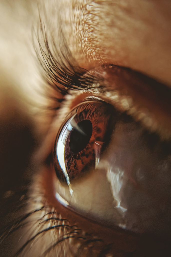
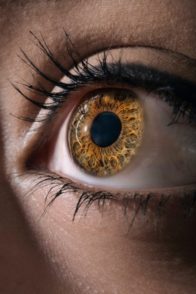
Michigan’s Premiere Ophthalmology Experts
Clear Vision Starts Here
With an unparalleled level of expertise, our doctors and staff provide the highest level of medical and surgical vision care to our patients.
Cataracts
A clouding of the lens of the eye or its surrounding transparent membrane that obstructs the passage of light. This cloudiness can cause a decrease in vision and may lead to eventual blindness.
Inside our eyes, situated behind the iris and pupil, is our crystalline lens. Typically, the lens is clear, but when a cataract develops, the lens becomes cloudy. Often, the vision will seem like you are looking through a dusty or dirty wind shield. Most people develop cataracts gradually due to the natural aging process, but cataracts can develop quicker from exposure to UV light, smoking, diabetes, eye injury, or the use of certain medication, such as steroids.
Symptoms of Cataracts
- Blurred vision
- Glare and halos around lights
- A feeling of “film” over the eye(s)
- Difficulty reading in ordinary light
- Sensitivity to bright lights
- Difficulty driving at night
- Double vision in one eye
- Bright colors look dull or faded “yellow or gray”
There are 3 types of cataracts:
Subcapsular Cataract: This type occurs at the rear of the lens. It’s most prevalent in people with diabetes or people who take high doses of steroid medications.
Nuclear Cataract: This type develops deep within the nucleus of the lens. Nuclear cataracts are the most common age-related cataracts.
Cortical Cataract: Characterized by whitish cloudy areas, these cataracts develop on the outside edge of the lens, called the cortex.
How are Cataracts treated?
The only way to remove a cataract is with surgery. At early stages of development, changes in eye glass prescription can temporally help with the symptoms of cataracts. Surgery should be considered when symptoms are keeping you from enjoying everyday life

Macular Degeneration
Progressive deterioration of a critical region of the retina called the macula. This disorder can lead to irreversible loss of central vision, although peripheral vision is retained. Vision may be gray, hazy or distorted in early stages.
Macular degeneration (AMD) affects the retina and damages your central vision. It affects the fine details centrally, but peripheral vision will stay unchanged. It is a leading cause of vision loss in people 50 years old or older. Many people don’t realize they have vision changes from AMD until their vision is compromised. Preventative eye health exams are an important tool to conserve vision and look for early signs of AMD before vision problems occur.
There are two forms of AMD: wet and dry. There is no treatment for dry AMD, but certain vitamins and minerals might help. Wet AMD can by treated with medication and/or laser surgery.
Amsler Grid: An Amsler Grid is a tool to detect any changes in how you are seeing. There are apps you can download or paper grids you can use as well. Follow your doctor’s instruction on how often to use the grid and follow up with scheduled appointments to help manage the effect of AMD.
A large study was conducted that revealed a certain combination of vitamins and minerals slow down the progression of AMD and your doctor can let you know if you would benefit from the vitamins and minerals
- Vitamin C (500mg)
- Vitamin E (400 IU)
- Lutein (10 mg)
- Zeaxanthin (2 mg)
- Zinc (80 mg)
- Copper (2 mg)
Flashes and Floaters
Flashes often look like lightning streaks or flashing lights in your vision. You may notice them for days or weeks at a time. Flashes happen when the vitreous rubs or pulls on your retina.
Floaters look like tiny specks or dots, lines or cobwebs in your vision. Floaters are tiny clumps of gel or cells inside the vitreous that fills your eye. You are actually noticing shadows of the clumps reflecting on the retina. They are often most visible when looking at something white or bright, like a piece of paper or the clear sky.
Symptoms can also be signs of a more serious condition. Flashes and/or Floaters can be symptoms of a torn or detached retina. So, if you notice flashes and/or floaters, you should have a dilated examination to determine if there is any concern.
- You notice a lot of new floaters
- You notice a lot of flashes
- A shadow appears in your peripheral (side) vision
- A gray curtain covers part of your vision

Refraction
Refraction is a testing procedure that measures how much optical (focusing) error an eye has. Certain eye measurements are taken using a variety of instruments. Based on these measurements, a series of trial lenses are placed in front of your eyes, and you are asked to compare one lens with another to determine which lens combination offers you better vision. This leads to a determination of how well you see and can be used to write a prescription for eyeglasses.
Why doesn't my health insurance pay for Refraction?
Most health insurance plans were not designated to pay for routine procedures. Medicare, Medicaid and most private commercial policies will not pay for refraction because it is considered routine.
Who has decided that Refraction is not covered?
It is our government (for Medicare and Medicaid) or your private commercial insurance company that determines exactly which services are covered, not your individual doctor.
What is Somerset’s policy?
In order to provide the very best eye care, refraction will be performed for all new patients, those presenting with decreased vision. Our office fee for a refraction is (NEED PRICE) and this fee is collected at the time of service in addition to any co-pay your health insurance plan may require.
We cannot file insurance on both the medical and routine vision plan for the same visit.
Diabetic Health Eye Exam
People with Diabetes need preventative eye health examinations, particularly those who have struggled with diabetes for years, can develop Diabetic Retinopathy. Diabetic Retinopathy is a degenerative eye disease that affects the retina.
Diabetes makes the body’s blood vessels especially susceptible to damage, particularly those in the kidneys and eyes. When the retinal eye blood vessels are damaged by diabetes, the blood and/or fluids leak into the eye. The retina may also form new, fragile blood vessels that can hemorrhage and leak as well. Either of these situations can lead to permanent vision loss.
Two Stages of Diabetic Eye Disease
NPDR (non-proliferative diabetic retinopathy) is the early stage of diabetic eye disease. With NPDR, tiny blood vessels leak or close off in the retina causing vison loss. It is the most common reason why people with diabetes lose their vision.
PDR (proliferative diabetic retinopathy) is the more advanced stage of diabetic eye disease. It occurs when the retina starts to grow blood vessels. The vessels grown are weak and fragile and often bleed easily. Scar tissue can form and cause problems that affect both central and peripheral (side) vision.

Dry Eyes
Our eyes need tears to stay comfortable and healthy. Tears help us see better as well. If your eyes do not make enough tears or they make the wrong type of tears, it is called Dry Eyes.
The tear film is made up of three layers:
- An oily layer – the outside layer that creates a smooth tear surface that keeps eye from drying out as quickly.
- A watery layer-the middle layer that cleans the eye and washes away debris or particles
- A mucous layer-the inner layer that assists in spreading the watery layer over the eye’s surface and help tears “stick” to the eye
Symptoms of Dry Eye
- Stinging and burning
- Scratchy or feeling like something is in your eye
- Red or irritated
- Unable to wear contacts comfortably
- Lots of tears or watery eyes
LASIK
The word LASIK is an acronym for “laser-assisted in situ keratomileusis.” It is a refractive procedure that reshapes the cornea to correct nearsightedness, farsightedness and astigmatism. LASIK surgery is considered a “refractive surgery” because its goal is to improve vision at distance without the need for glasses or contact lenses.
Benefits to LASIK surgery include little or no post-operative discomfort, immediate vision improvement, and the ability to drive or return to work quicklysometimes as soon as the next day. Most patients require no corrective eyewear after surgery although patients over 40 may require reading glasses.
LASIK is the most common refractive surgery performed today.
If LASIK is not an option, there are other “refractive surgeries” available to make you independent of glasses or contacts.

Additional Eye Conditions
Glaucoma
Any group of eye diseases characterized by abnormally high intraocular fluid pressure. This can lead to a damaged optic disk, hardening of the eyeball and partial to complete loss of vision.
Strabismus
A condition in which the eyes do not point in the same direction.
Retinal Detachment
A separation of the retina from the retinal pigment epithelium in the back of the eye. In most cases retinal detachment develops slowly.